Tuesday, December 14, 2010

Man Officially Cured of HIV

Source

Man Officially Cured of HIV


For the first time, a man has been declared officially cured of HIV. The remedy may nearly have killed him, but it opens a door—just a crack—to hope that we may someday kill off the scourge for good.

Strangely enough, the diagnosis that most concerned Timothy Ray Brown in 2007 was acute myeloid leukemia. HIV has been increasingly thought of as a manageable disease, though certainly a terribly burdensome one. What brought the 42-year old Brown under the care of Germany's Charite Universitatsmedizin Berlin hospital was the more immediate threat his cancer posed.

The treatment Brown underwent was aggressive: chemotherapy that destroyed the majority of his immune cells. Total body irradiation. Finally, a risky stem-cell transplant that nearly a third of patients don't survive—but that appears to have completely cured Brown of HIV.

Doctors were savvy when they chose a stem cell donor for Brown. The man whose bone marrow they used has a particular genetic mutation, present in an incredibly small percentage of people, that makes him almost invulnerable to HIV. With Brown's own defenses decimated by treatments, the healthy, HIV-resistant donor cells repopulated his immune system. The initial indications that the virus had abated were promising. But only just now, having taken no antiretroviral drugs since the transplant, and following extensive testing shows no signs whatsoever of HIV, have his doctors given the official word:

He's cured.

What does this mean for the future of treatment? It's not as though every HIV patient can or would want to go through the tremendous suffering that was prelude to Brown's recovery, or be able to afford the procedure if they could or did. But for the first time, we know that HIV can be cured, not just managed. It opens new avenues of research—gene therapy, stem cell treatments—that may otherwise have been thought dead ends. [AIDS Map]

Tuesday, November 30, 2010

Reversing Aging

Harvard team successfully reverses the aging process in mice

By Loz Blain

23:04 November 29, 2010

Chromosomes, with their telomere caps highlighted. Looking after these telomeres could be ...

Chromosomes, with their telomere caps highlighted. Looking after these telomeres could be the key to longer life.

The aging process - it's undignified, unwanted, and many would say unnecessary. After all, the cells in your body are constantly replacing themselves – why can't they do it without causing progressive degradation of organs that lead to discomfort, weakness and death? Well, perhaps they can. Harvard scientists have discovered that by controlling certain genetic processes in mice, they can not only slow down the aging process, but "dramatically" reverse it throughout the body. It's a massive discovery, but it won't be able to be used in humans yet without some pretty scary consequences.

Telomeres

The Harvard study focused on part of the cell division process called 'telomere shortening.' If you picture a chromosome as an X-shaped unit of DNA, the telomeres are the little caps at the end of each strand.

As cells divide, their DNA splits in half to form two new cells – but a bit of genetic information is lost at the end of each strand with each division. That's what telomeres are for – they contain a bunch of useless DNA that acts as a buffer zone so that no important DNA is lost from our chromosomes. Gradually, over time, the telomere erodes away to a level where each cell division actually starts destroying bits of important DNA – and this gets to a point where the cell can no longer reproduce itself. You can imagine what that starts doing to bodily organs as time goes by.

So in a way, telomeres are there as a built-in limit to how many times a cell can divide itself – they're part of the built-in biological clock that causes aging, body deterioration and death.

The Harvard Study

And that's where this recent study was focused. Ronald DePinho and a team of Harvard colleagues experimented on mice to see what happens when steps are taken to stop telomeres from shortening.

The group looked at the enzyme telomerase, which can replenish the telomere after replication and effectively lengthen it so that a cell can live for longer.

They bred a group of genetically-engineered mice that lacked the ability to produce telomerase – and watched as these mice showed rapid and very early onset symptoms of aging.

Then, they gave the mice injections to re-activate the telomerase enzyme – expecting to see the aging process slow down to normal levels. Instead, they watched in astonishment as the mice appeared to age backwards, their withered organs repairing themselves even to the point of new neurons beginning to sprout in their brains.

In essence, repairing the telomeres seemed to be able to reverse the aging process and make the mice physiologically younger, despite already suffering the ravages of age.

But it's not so simple for humans

While this study was a huge step forward in our understanding of how telomeres and telomerase impact the aging process, there's a big issue preventing this sort of treatment from going straight into human testing.

Mice produce telomerase all throughout their short lives, but the enzyme is switched off in adult humans with our longer life spans, because when our cells are allowed to divide and replicate unchecked, they have a nasty habit of developing into cancers.

Although a telomerase-activating compound was recently discovered, telomerase rejuvenation in adults is directly linked to the development and spread of cancers throughout the body – so while a telomerase-based anti-aging treatment might produce valuable results in older humans, scientists also believe it would kick the risk of cancer up several orders of magnitude.

So the study isn't the sort of breakthrough that will lead directly and immediately to any sort of treatment for humans – but it's another step forward in science's inevitable march towards human immortality. Imagine what a gift it would be for an 80-year-old to be given an injection that could tell the cells in his body to rebuild itself in the physical health he enjoyed as a 25-year-old, knowing everything he knows.

There are already debates flaring over the ethics of extending lifespan, and the question of whether humans should submit themselves to the long-accepted ravages of aging and death or use our amazing command of science to avoid them. But the fact is, we're going to work out how to do it sooner or later, whether or not it involves this sort of telomere repair process, and a significant number of people will want to use such a technology to repair their bodies and see what life is like in the next few centuries.

Would you be one of them?

Monday, November 22, 2010

Reasons Why We Will Eventually Reverse Aging - Aubrey de Grey

One day, our species may look back at these times and gasp at how we once aged and slowly died.


Excerpt from the following: http://www.slate.com/id/2274582/pagenum/all/

The classical areas of regenerative medicine have been moving ahead at an unprecedented pace in recent years. Some of the highlights have, with good reason, been celebrated in the mainstream media: The foremost example is the development by Shinya Yamanaka, and subsequent refinement by numerous groups, of a method to convert adult cells into a state very similar to embryonic stem cells. These cells, termed "induced pluripotent stem cells" or iPS cells, appear to have almost all the versatility of true embryonic stem cells, but can be obtained far more easily, in far greater numbers, and without the main ethical challenges that have confronted the embryonic stem cell field. Stem Cells

Less celebrated, but possibly of similar significance, is an advance in tissue engineering. Following the pioneering work of Doris Taylor, many groups are now pursuing a creative solution to the main problem that has always dogged tissue engineering: vascularization. The blood supply is a vital component of any solid organ, and it has proved hugely difficult to cajole the body to create a competent vasculature in an artificial organ before the cells in that organ have succumbed to deprivation of oxygen or nutrients. Taylor's solution is to "decellularize" an organ from another individual—possibly not even from a human—leaving only the extracellular matrix that defines the vasculature, and then to repopulate this scaffold with suitable cells typically taken from the intended recipient of the organ. This achieves the main goal of tissue engineering, the rejection-free replacement of a damaged organ. Replacing Damaged Organs

Less conspicuous is "molecular regenerative medicine"— repair of the intracellular structure of live cells in situ, or of extracellular structures, as opposed to the wholesale replacement of cells or organs. One of the most impressive advances within this field relates to Alzheimer's disease.The amyloid plaques that accumulate in the brains of Alzheimer's sufferers can be removed by vaccination, a trick that was first demonstrated (in mice) a decade ago. Doubts surfaced thereafter concerning whether this could work in humans, but the approach looks promising at present, with clinical trials having progressed to Phase III. Removal of plaques is unlikely to constitute a complete cure for Alzheimer's disease, but it is almost certain to be part of an eventual combination treatment.Removing Dead and Diseased Cells

At an earlier stage, but still immensely exciting, is the corresponding treatment for "molecular garbage" that accumulates not between cells but inside them. This is a more challenging aspect of aging, because the material in question is resistant to breakdown by any human enzymes. The solution being pursued by the SENS Foundation, the charity of which I am the chief science officer (SENS stands for Strategies for Engineered Negligible Senescence), is to identify non-human enzymes that can do the job and introduce them into our cells. The biggest initial challenge is to identify such enzymes, but we have now done that with respect to the main types of garbage responsible for cardiovascular disease and macular degeneration. This has already led to several peer-reviewed academic publications, and we hope to bring it to the stage of testing in mice within the near future. Eliminating Molecular Garbage

Additional targets being pursued by SENS Foundation include the elimination of cells that refuse to die when the body wants them to—a major aspect of the decline of the immune system with age, among other things—and the prevention of mitochondrial mutations by putting modified copies of the mitochondrial DNA into the nucleus. These projects are moving forward with impressive speed, though slowed by resource limitations.

When all these components are combined, will we have bona fide rejuvenation biotech? We won't know that for certain until we complete the development of all those components, at least in mice. But every type of "aging damage" of which we're aware—and for which SENS Foundation and others are developing interventions—has been known about for well over a quarter of a century. If some other factor is at work in aging, then biologists should have identified it by now. It looks as though all that stands between us and control of aging is hard work. Let's up the pace.

Monday, November 8, 2010

Blood created from Skin

Personalizing production of blood would reduce likelihood of rejection

Canadian scientists have transformed pinches of human skin into petri dishes of human blood -a major medical breakthrough that could yield new sources of blood for transfusions after cancer treatments or surgery.

The discovery, by researchers at McMaster University in Hamilton, Ont., could one day potentially allow anyone needing blood after multiple rounds of surgery or chemotherapy, or for blood disorders such as anemia, to have a backup supply of blood created from a tiny patch of their own skin -eliminating the risk of their body's immune system rejecting blood from a donor. Researchers predict the lab-grown blood could be ready for testing in humans within two years.

The achievement, published yesterday in the journal Nature, raises the possibility of personalizing blood production for patients for the first time.

"This is a very important discovery. I think it represents a seminal contribution" to the rapidly evolving field of stem-cell research, said Michael Rudnicki, scientific director of the Canadian Stem Cell Network and director of the Regenerative Medicine Program at the Ottawa Hospital Research Institute.

"That one can play with the fate of a cell and force it sideways into something that it doesn't at all resemble, and then being able to use it, is tremendously exciting."

The procedure is also relatively simple. It involves taking a small piece of skin, extracting fibroblasts - abundant cells in the skin that make up the connective tissue and give skin its flexibility -and bathing them in growth factors in a petri dish. Next, by adding a single protein that binds to DNA and acts as an on/off switch, the researchers turned on or off 2,000 genes and reprogrammed the skin cells to differentiate or morph into millions of blood progenitors -the cells the produce blood.

They generated multiple different blood-cell types - oxygen-ferrying red blood cells, infection-fighting white blood cells, cells that make platelets needed for healing, and macrophages, the garbage trucks of the blood system that swallow and break down foreign material.

The work was repeated several times over two years using skin from adults, as well as neonatal foreskin, demonstrating, according to background material, that it could work "for any age of person."

And while other researchers have reprogrammed fibroblasts into neurons, cardiac cells and even macrophage-like cells in mice, the McMaster team converted skin directly to blood using human skin.

The first to benefit could be patients with leukemia, whose blood undergoes genetic changes that turn it cancerous and who often need-bone marrow transplants, or those with lymphomas, such as Hodgkin's disease.

Bone marrow contains stem cells that produce blood cells. If the bone marrow is coming from a donor, "first of all, you have to find a match, which can often be a problem, especially for smaller ethnic groups," says Christine Williams, director of research at the Canadian Cancer Society Research Institute. There's also a risk of rejection, where the body sees the matched cells as foreign.

Using a patient's own bone marrow gets around the rejection risk. "But if you take the patient's own blood stem cells and put them back in after radiation, you're often re-transplanting cells that have the same mutation as the tumour," Williams says.

The skin cells of leukemia patients "don't have any genetic abnormalities," says Mick Bhatia, leader of the McMaster team. "So, if we could take the skin cells and generate blood, we could use that to transplant leukemic patients themselves."

The finding also has the potential of "making bone marrow transplant HLA (tissue) matching and paucity of donors a thing of the past," said Alain Beaudet, president of the Canadian Institutes of Health Research, which helped fund the study.


Tuesday, November 2, 2010

Cure for the Common Cold?

A cure for the common cold may finally be achieved as a result of a remarkable discovery in a Cambridge laboratory

By Steve Connor, Science Editor

In a dramatic breakthrough that could affect millions of lives, scientists have been able to show for the first time that the body's immune defences can destroy the common cold virus after it has actually invaded the inner sanctum of a human cell, a feat that was believed until now to be impossible.

The discovery opens the door to the development of a new class of antiviral drugs that work by enhancing this natural virus-killing machinery of the cell. Scientists believe the first clinical trials of new drugs based on the findings could begin within two to five years.

The researchers said that many other viruses responsible for a range of diseases could also be targeted by the new approach. They include the norovirus, which causes winter vomiting, and rotavirus, which results in severe diarrhoea and kills thousands of children in developing countries.


Viruses are still mankind's biggest killers, responsible for twice as many deaths as cancer, essentially because they can get inside cells where they can hide away from the body's immune defences and the powerful antibiotic drugs that have proved invaluable against bacterial infections.

However, a study by a team of researchers from the world-famous Laboratory of Molecular Biology in Cambridge has shown that this textbook explanation of the limits of the human immune system is wrong because anti-viral antibodies can in fact enter the cell with the invading virus where they are able to trigger the rapid destruction of the foreign invader.

"In any immunology textbook you will read that once a virus makes it into a cell, that is game over because the cell is now infected. At that point there is nothing the immune response can do other than kill that cell," said Leo James, who led the research team.

But studies at the Medical Research Council's laboratory have found that the antibodies produced by the immune system, which recognise and attack invading viruses, actually ride piggyback into the inside of a cell with the invading virus.

Once inside the cell, the presence of the antibody is recognised by a naturally occurring protein in the cell called TRIM21 which in turn activates a powerful virus-crushing machinery that can eliminate the virus within two hours – long before it has the chance to hijack the cell to start making its own viral proteins. "This is the last opportunity a cell gets because after that it gets infected and there is nothing else the body can do but kill the cell," Dr James said.

"The antibody is attached to the virus and when the virus gets sucked inside the cell, the antibody stays attached, there is nothing in that process to make the antibody to fall off.

"The great thing about it is that there shouldn't be anything attached to antibodies in the cell, so that anything that is attached to the antibody is recognised as foreign and destroyed."

In the past, it was thought that the antibodies of the immune system worked entirely outside the cells, in the blood and other extra-cellular fluids of the body. Now scientists realise that there is another layer of defence inside the cells where it might be possible to enhance the natural anti-virus machinery of the body.

"The beauty of it is that for every infection event, for every time a virus enters a cell, it is also an opportunity for the antibody in the cells to take the virus out," Dr James said.

"That is the key concept that is different from how we think about immunity. At the moment we think of professional immune cells such as T-cells [white blood cells] that patrol the body and if they find anything they kill it.

"This system is more like an ambush because the virus has to go into the cell at some point and every time they do this, this immune mechanism has a chance of taking it out," he explained.

"It's certainly a very fast process. We've shown that once it enters the cell it gets degraded within an hour or two hours, that's very fast," he added.

The study, published in the journal Proceedings of the National Academy of Sciences, involved human cells cultured in the laboratory and will need to be replicated by further research on animals before the first clinical trials with humans.

One possibility is that the protein TRIM21 could be used in a nasal spray to combat the many types of viruses that cause the common cold. "The kind of viruses that are susceptible to this are the rhinoviruses, which cause the common cold, noravirus, which causes winter vomiting, rotavirus, which cause gastroenteritis. In this country these are the kind of viruses that people are most likely to be exposed to," Dr James said.

"This is a way of boosting all the antibodies you'd be naturally making against the virus. The advantage is that you can use that one drug against potentially lots of viral infections."

"We can think of administering these drugs as nasal sprays and inhalers rather than taking pills... It could lead to an effective treatment for the common cold," he said. "The beauty of this system is that you give the virus no chance to make its own proteins to fight back. It is a way for the cell to get rid of the virus and stay alive itself."

Sir Greg Winter, deputy director of the MRC Laboratory of Molecular Biology, said: "Antibodies are formidable molecular war machines; it now appears that they can continue to attack viruses within cells. This research is not only a leap in our understanding of how and where antibodies work, but more generally in our understanding of immunity and infection."

How the virus is tackled

* 1 Virus (purple) circulating in the bloodstream recognised by antibodies (yellow) of the immune system

* 2 Virus attaches to outer cell membrane with antibodies still attached

* 3 Virus invades the cell membrane and emerges inside the cell

* 4 Remains of cell membrane disappear and the virus is free to hijack the cell

* 5 TRIM21 protein (blue) recognises attached antibodies as foreign material

* 6 Powerful virus-destroying machines (cylinders) attracted to virus by TRIM21

* 7 Virus rapidly broken down and disabled within hours

Friday, October 22, 2010

HIV Cured by Immune System Stimulation


International activists are lashing out at world leaders for failing in their promises to do more to treat people with HIV. The criticism comes during the AIDS conference which is underway in Vienna, calling for an end to 'treatment discrimination'. Dr Marco Ruggiero, from the University of Florence in Italy, supports the view that the public has been misinformed about HIV.

Now, maybe these are the alternate "quacks". Well the scientist who discovered HIV agrees with them

Killing Leukemia


It used to be a disease that very few children could survive. But thanks to a breakthrough by a pioneering clinic in Russia, 70 per cent of young sufferers of leukemia now make a full recovery. Karina Falakhutdinova is seven years old -- but if it had not been for the pioneering new treatment she would not have seen her first birthday.

Regenerating body parts

Woman's persistence pays off in regenerated fingertip

By Elizabeth Cohen, CNN Senior Medical Correspondent
September 9, 2010 -- Updated 2051 GMT (0451 HKT)
When the slam of a door chopped off the tip of her pinky, doctors told Deepa Kulkarni nothing could be done. She did her own research and proved them wrong.
STORY HIGHLIGHTS
  • Sometimes doctors don't know about new treatments being used by other physicians
  • Don't hesitate to e-mail a doctor who's a leader in the field that could help you
  • That treatment that looks perfect on the internet might work for you -- but it might not

(CNN) -- After running inside from a rainstorm one Friday evening last January, Deepa Kulkarni leaned against the doorway with her right hand to take off her boots. Then, in an effort to make sure the dog didn't get out, someone slammed the door hard, and it landed right on her pinky.

Kulkarni thought the door had only bruised her finger, but then she looked down and saw the tip of her pinky lying on the floor.

"I swooped down and picked it up before the dog got it," she remembers. "At first I was fine, but then I saw the blood -- there was so much blood -- and I felt woozy."

Her husband, Ajit, called an ambulance, and as soon as his parents arrived to take care of their two young children, Kulkarni retrieved her pinky tip from the freezer -- it had broken off just above the base of her fingernail -- and they headed from their home in Davis, California, to the emergency room.

Video: Woman grows back finger tip

That trip to the hospital was the beginning of a five-week odyssey -- one that involved defying doctors, e-mailing specialists around the country and -- in her words -- being pushy and demanding in order to make her pinky whole again.

'100 percent chance of failure'

"The doctor who was on call at the emergency room told me there was no way he could reattach my pinky," she says. "I didn't like that, so I asked to see a specialist."

An orthopedic surgeon concurred with the ER doctor, and made an additional recommendation: He'd have to amputate even more of the finger so it would heal properly.

"I was like, no way, with so much technology out there, there must be some other way to do this," she said. "But he said he wouldn't even attempt to reattach it. He said was there was a 100 percent chance of failure," she remembers.

Disappointed but defiant, Kulkarni declined the surgeon's treatment, and the doctors sent her home with a bandaged finger and painkillers.

'I am writing to you out of desperation.'

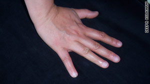
Deepa Kulkarni displays her hand after the accident.

Arriving home at midnight, Kulkarni fell asleep, and the next morning immediately got on her computer. With her nine functioning fingers she searched for hope -- any hope -- that her pinky could be repaired.

Kulkarni, a former human resources manager who is now in law school full time, was accustomed to doing research, but after searching for hours, she still couldn't find any new ideas for reattachment.

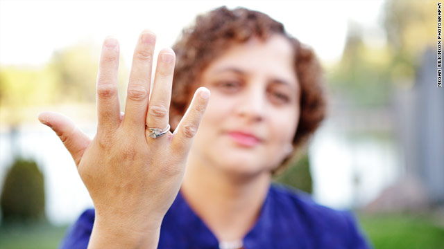
Kulkarni's pinky grew back after she persuaded a doctor to administer a procedure she had read about on the internet.

She did, however, find something else: stories on "60 Minutes" and "Oprah" about a relatively new procedure called tissue regeneration, which had made amputated finger parts grow back for other patients.

She went to the website for Dr. Stephen Badylak, the University of Pittsburgh physician who helped pioneer the procedure. A little more searching revealed his e-mail address.

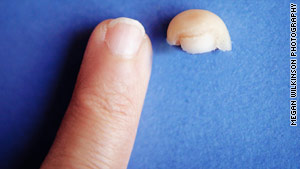
Kulkarni displays her new pinky next to the tip of her old pinky.

"Dear Dr. Badylak," she began. "I am writing to you out of desperation...I ran across your article on the internet and decided to write to you. Can you tell me if [tissue regeneration] works and what my options are to get the treatment?"

She heard back the next day, a Sunday, from one of Dr. Badylak's colleagues. He gave her more details about the procedure, and some bad news: He didn't know of any doctors near her using regenerative medicine.

She e-mailed him photos of her damaged pinky, and he said the team of doctors in Pittsburgh would review them and decide whether they could help her. In the meantime, Kulkarni sent out an all-call in search of a physician near them who used tissue regeneration.

"I e-mailed people at UCLA and Berkeley, and my husband and I called everyone we knew who was a doctor or might know a doctor," she says.

Still, no one had any ideas for her. One friend introduced her to Dr. Malathi Srinivasan, an internist at the University of California-Davis Medical Center, who called Kulkarni from her cell phone while driving home from a skiing trip at Lake Tahoe.

Srinivasan didn't know any doctors with experience in tissue regeneration, but she did give her something Kulkarni badly needed at this point: hope.

"I encouraged her to keep looking, and I did some PubMed searches myself," Srinivasan says, referring to the National Library of Medicine's online collection of research materials.

Seven weeks later, a new pinky

Eventually, Kulkarni made an appointment with Dr. Michael Peterson, an orthopedic surgeon in Davis. At first, Kulkarni says he was hesitant to try tissue regeneration since he hadn't done it before, but she gave him some research materials, and she says eventually he agreed to try it.

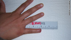
The therapy involved cleaning out the finger and removing scar tissue -- a process called debridement -- and then dipping her finger into MatriStem wound powder. After seven weeks of treatment, her fingertip grew back (as shown in the before and after photos above).

"Even now it's not perfect. It's shorter than the other pinky, but just by looking at it you can't tell it was an amputated finger," she says. "I'm able to do everything I could do before. I wash dishes. I cook," she says, adding that she's had physical therapy to decrease tingling in her finger caused by severed nerves.

Kulkarni still keeps her severed pinky tip in the freezer. She thawed it out so CNN could take a photograph of the pinky tip alongside her regenerated finger.

Five lessons learned

Here are lessons Kulkarni learned through her pinky ordeal, plus a few from the doctors who worked with her:

1. Realize your doctor doesn't know everything

"I grew up in India, and doctors there were looked up to as being next to God, and they knew everything and could cure you," Kulkarni says. "What I found out is that even though I like my doctors, I don't have to take every recommendation they give me. I can do my own research."

Srinivasan, the physician who encouraged Kulkarni not to give up, says sometimes doctors don't know about new, cutting-edge treatments being used by other physicians.

"Some doctors are more out-of-date or up-to-date than others," says Srinivasan, an associate professor of medicine at UC-Davis Medical Center who does research on shared decision-making between doctors and patients.

Kulkarni said even when her prospects looked dim, she never completely gave up hope.

"My husband and I are quite pushy, and my husband is smart -- he has a Ph.D. in electrical engineering. We weren't going to take no for an answer," she says.

Petersen, Kulkarni's surgeon, says he's learned from many patients who've come to him having done research on the internet.

"I get a lot of good ideas from patients," he says. "One of the things we have to do as physicians is to listen to our patients."

2. E-mail doctors you don't know

Even though you're a stranger, if you write to a doctor who's a leader in the field, you might get an e-mail in return.

"The doctor from Pittsburgh wrote me back the very next day, on a Sunday," Kulkarni says. "I was so surprised. Maybe I sounded really desperate."

Even if you can't afford to fly out to the expert for treatment, he or she might be able to lead you to a local doctor who can help.

3. Don't be afraid to be a 'bad' patient

Kulkarni said she knows the doctors she saw in the emergency room might have perceived her as an annoying patient when she rejected their treatment options.

"I didn't care," she says. "Imagine if I hadn't pursued other options because I was worried about what other people think -- I didn't want to live with that regret."

4. Be organized

Be fastidious about keeping records. Kulkarni, for example, photographed her pinky at various stages, and she e-mailed the images to various doctors and later to the insurance company to convince them to reimburse her the $1,665 she spent on the drug that regenerated her pinky.

5. Recognize you might be wrong

That treatment that looks perfect on the internet might work for you -- but it might not.

"Sometimes patients are pretty certain through their own research what their problem is and what they need," says Petersen. "It's not up to us as doctors to agree with them. Sometimes part of my role is to guide them that, in reality, that isn't your problem and I don't recommend that treatment."

Learn more about Elizabeth Cohen's book on being an Empowered Patient. Video

CNN's Sabriya Rice contributed to this report.

Popular Posts